
Keratoconus: Definition & Overview
Keratoconus, pronounced (ker-uh-toe-KOH-nus), is a chronic, degenerative disease of the cornea where it starts thinning and bulges out into a cone shape as opposed to its normal round shape. The adjacent illustration shows a side view of a normal, round-shaped cornea versus a cone-shaped, keratoconus cornea. The explainer-type video below provides information that explains everything about keratoconus. We also have a short summary of how we help our patients with keratoconus.

How Keratoconus Affects the Cornea
The cornea is the clear tissue covering the front of the eye, both the iris and the pupil. Its main purpose is to direct light onto the retina for clear vision. As the cornea weakens, it can no longer maintain its normal curvature against the internal pressure of the eye.
Thus, the cornea bulges out, causing astigmatism. This change in structure affects the person’s eyesight, so that images start blurring, which occurs at all distances, causing myopia (nearsightedness) as well as hyperopia (farsightedness).
The adjacent photograph shows a close-up view of a man’s eye with keratoconus. Note the obvious cone-shaped cornea of the patient.
Living with Keratoconus: What You Should Know
Keratoconus only affects about 1 in 2000 people, so the rate of disease is low. The current US population is about 330 million, so by calculation, the US has about 165,000 keratoconus sufferers. But its effects can be devastating as it can cause blindness. It typically affects both eyes; however, it usually does not affect them to the same degree.
Here is a short news video about Golden State Warriors’ NBA superstar Steph Curry’s battle with keratoconus. Since he started wearing scleral contact lenses, his game has gotten significantly better, if you can believe that! It also shows what keratoconus is and how it affects your eyesight.

What Causes Keratoconus? Risk Factors to Know
Although a significant number of studies have been performed, the definitive cause of keratoconus is still not well understood. The risk factors given below are related to a predisposition for keratoconus.
For those individuals with keratoconus, it typically starts just after childhood, during puberty, in adolescence, and can progress until young adulthood, about 25-35 years old. Although in some people it never becomes progressive, so in that way, it’s an enigma.
There is a genetic component, as about 10% of the cases have at least one parent who also has the disease. A family history of Down Syndrome and asthma also increases your risk.
Those whose eyes are continually inflamed are more susceptible to keratoconus. The inflammation causes irritation and pain, resulting in eye rubbing. Chronic eye rubbing is thought to be a major cause of keratoconus progression. The image below shows a patient's inflamed eye exacerbated by eye rubbing.
Keratoconus is also associated with eye injuries, eye allergies, and connective tissue disorders such as Marfan syndrome and Ehlers-Danlos syndrome. Other eye diseases like retinitis pigmentosa increase the risk as well.
Can Keratoconus Be Prevented or Treated?
Unfortunately, at this time, we do not know how to prevent keratoconus, and there is no known cure. In addition, in the early stages, some people may not realize that they have it.
This makes it incredibly important to make an appointment for an eye examination with your eye doctor for early diagnosis and treatment of keratoconus.


Common Symptoms of Keratoconus
If you have one or more of the symptoms listed to your right, contact your optometrist at Good Eye Optometry to schedule a comprehensive eye examination to determine the cause. It may not be significant, but it’s a good idea to find out sooner rather than later.
- When you look at lights, you see halos or glare around them
- You develop a sensitivity to bright lights
- Your night vision is impaired
- Your vision becomes distorted or blurred
- Your eyeglasses or contact lenses prescription changes frequently
- Your vision suddenly gets worse, or your vision cloud overs
How Keratoconus Is Diagnosed
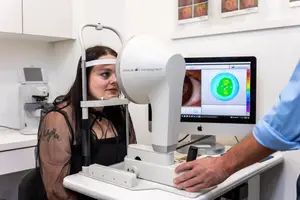
All eye care tests should be preceded by a thorough medical history and then a comprehensive eye exam to determine visual acuity and detect potential eye abnormalities. There are a couple of ways to diagnose keratoconus. The most accurate tool with the latest technology is a corneal topography machine, in which a computer-assisted 3D map of your cornea is created from thousands of digital measurements.
This method is non-invasive, painless, and only takes a few minutes while you stare into a concave cone. The corneal topography machine is shown in the adjacent photograph, where the test is about to begin.
A sample image of the results from the corneal topography test is shown in the image on your left, where the varying thickness of the cornea is color-coded.
These results are from a patient with severe keratoconus, as shown by the large red area indicating the steepness of the cornea in that area. This is explained in detail in the short video on corneal topography below.
Primary Tests for Corneal Irregularities
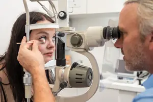
A slit-lamp machine can detect corneal anomalies by measuring the middle and outer layers of the cornea. So it can also diagnose keratoconus. In this test, your eye doctor shines a bright light into your eye while looking through a microscope to evaluate your cornea. It is both non-invasive and painless.
While there are other methods to measure your cornea, these are the primary tests.
Treatment Options for Keratoconus
We do know that treatment for keratoconus is essential so that it does not result in permanent vision loss. The treatment goals are correcting vision and minimizing disease progression. Treatment is determined by the stage of progression of the disease: early, intermediate, or advanced.
In the very early stages of keratoconus, people may not even know that they have the disorder. This is one reason why regular periodic eye exams are highly recommended. When keratoconus is first diagnosed in its early stage, the strategy is to correct your vision and obtain baseline data so we can determine its progression over time. At this point, your doctors may just order eyeglasses or a soft contact lens prescription to increase visual acuity.
Scleral Contact Lenses
If your keratoconus progresses to the intermediate stage, eyeglasses and soft contact lenses probably can no longer correct your vision. Scleral contact lenses are a great option that are custom-made to fit each patient’s eye. Therefore, they are more expensive than normal contact lenses. Scleral lenses are hard, gas-permeable contacts with a large diameter, so they can vault over the cornea and rest on the sclera, the white part of the eye. Even though large, scleral lenses are quite comfortable because the sclera has many fewer nerve endings when compared to the cornea. The area between the inside of the contact and the cornea is filled with a saline solution that helps with vision correction. The video below shows an interview with patient, Larissa, who explains that her scleral contact lenses are a godsend.
Collagen Cross-Linking
The great news is that the cornea can be strengthened by a relatively new process called collagen cross-linking or corneal cross-linking. The procedure was approved by the FDA in 2016 after clinical trials proved its efficacy. It is the only technique that has been shown to halt or slow keratoconus progression. In some cases, it improves the curvature of the cornea and becomes more normally shaped as it strengthens. Vision improves in many patients who undergo this procedure. Collagen, a protein, is an important component of the middle section of the cornea, which adds strength and flexibility to the cornea. In keratoconus, some of the collagen in the cornea is lost, causing weakness. This one-time procedure is performed by an ophthalmologist in the office, where the eye is bathed in a Vitamin B2 (riboflavin) solution while ultraviolet light activates the collagen to cross-link to other collagen molecules by forming strong chemical bonds. In the news interview below, Dr. Ryan Turner explains how this technique is being used to help keratoconus patients. In the next video, Dr. Terrence Doherty of Loden Vision Centers in Nashville, TN, walks you through a collagen cross-linking procedure with patient Michael.
If keratoconus reaches the advanced stage, you have two basic options, both of which involve eye surgery performed by an ophthalmologist.
Corneal Ring Implant
If your keratoconus becomes severe and contact lenses are causing too much pain, you may be a candidate for corneal ring surgery. Plastic, C-shaped rings are surgically inserted into the middle layer of the cornea to add strength, structure, and flatten the cornea. Other common names for these rings are INTACS® or ICRS. The procedure takes place in an outpatient surgical center. It’s quick, about 15 minutes per eye, simple, and painless. Vision correction typically is quite dramatic as a patient goes from unable to read to almost everyone can pass the driver’s license eye exam without eyeglasses, and about half the patients have 20/20 vision.
Corneal Transplant
A corneal transplant may be necessary if your keratoconus progresses to the advanced stage, and you are not a candidate for a corneal ring implant. For example, if your cornea has scarring or traumatic injury. The surgery usually takes place as an outpatient and takes about an hour per eye. Your cornea is excised and replaced with a donor cornea. The biggest issue is preventing tissue rejection, but despite this, it has a 95% success rate, one of the highest success rates of all transplant surgeries. In most cases, the patient will still require corrective lenses even after a transplant.


Next Steps if You’re Noticing Symptoms
If you think you are experiencing any of the signs or symptoms of keratoconus, please make an appointment with us at Good Eye Optometry. Our optometrists have extensive experience working with and successfully managing patients with keratoconus. Remember, early diagnosis is very important so we can monitor its progression and recommend the proper treatment.
For more information on keratoconus, go to:
The National Keratoconus Foundation